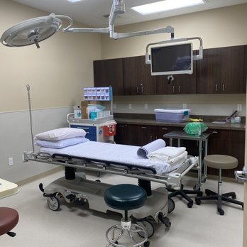
Direct Imaging PC Photos


You might also consider


More like Direct Imaging PC
Recommended Reviews - Direct Imaging PC



More info about Direct Imaging PC
6101 Summitview Ave
Ste 200
Yakima, WA 98908
Directions
(509) 902-8857
Call Now
Visit Website
http://www.directimagingyakima.com
Hours
Payment methods
Other Amenities
From the Business
Ask the Community - Direct Imaging PC












Why does Yelp recommend reviews?
3 reviews that are not currently recommended
The reviews below are not factored into the business's overall star rating.

3 years ago
3 years ago
2 years ago
Direct Imaging PC - diagnosticimaging - Updated August 2026
Browse Nearby
BEST of Yakima, Washington Restaurants near Direct Imaging PC
BEST of Yakima, Washington Coffee near Direct Imaging PC
BEST of Yakima, Washington Things to Do near Direct Imaging PC
BEST of Yakima, Washington Sushi near Direct Imaging PC
BEST of Yakima, Washington Family Practice near Direct Imaging PC
BEST of Yakima, Washington Optometrists near Direct Imaging PC
BEST of Yakima, Washington Nurseries & Gardening near Direct Imaging PC
BEST of Yakima, Washington Thrift Stores near Direct Imaging PC
Browse Brands
Related Searches in Yakima, WA
Accurate Laboratory Testing Available in Yakima, Washington
Best Imaging Centers Around Yakima, Washington
Best Pediatricians Practicing in Yakima, Washington
Browse Expert Sleep Specialists in Yakima, Washington
Consult a Skilled Neurologist Near Yakima, Washington
Effective Pain Management Services in Yakima, Washington
Experienced Family Doctor Care Available in Yakima, Washington
Experienced Nurse Practitioner Care in Yakima, Washington
Experienced Obstetricians & Gynecologists in Yakima, Washington
Experienced Podiatrists Serving Patients in Yakima, Washington
Experienced Primary Care Physician in Yakima, Washington
Experienced Psychiatrists Near Yakima, Washington
Experienced Radiologists Serving Yakima, Washington
Experienced Rheumatologists Practicing in Yakima, Washington
Explore Medical Centers in Yakima, Washington
Explore Weight Loss Centers Around Yakima, Washington
Explore the Best Hospitals Available in Yakima, Washington
Family Doctor Accepting New Patients in Yakima, Washington
Find Qualified Osteopathic Physicians Practicing in Yakima, Washington
Find Top Emergency Rooms Available in Yakima, Washington
Find Top-Rated Dermatologists in Yakima, Washington
Find Top-Rated Physical Therapy Services in Yakima, Washington
Find Top-Rated Sports Medicine Services in Yakima, Washington
Find Trusted Colonoscopy Services in Yakima, Washington
Find Trusted Orthopedists in Yakima, Washington for Expert Care
Get Open MRI Services Available in Yakima, Washington
Leading Ultrasound Imaging Centers in Yakima, Washington
Quality Diagnostic Services Available in Yakima, Washington
Quality Health & Medical Services in Yakima, Washington
Reliable Family Practice Services in Yakima, Washington
Reliable MRI Imaging Centers in Yakima, Washington
Schedule With a Primary Care Doctor in Yakima, Washington
Schedule With an Orthopedic Doctor in Yakima, Washington
Schedule a Mammogram Screening in Yakima, Washington
Top Female Doctors Available in Yakima, Washington
Top Hand Specialist Care Available in Yakima, Washington
Top-Rated Blood Labs for Testing in Yakima, Washington
Top-Rated Chiropractors Available in Yakima, Washington
Top-Rated Clinic Services Available in Yakima, Washington
Top-Rated Spine Surgeons Available in Yakima, Washington
Trusted Gynecologist Care Providers in Yakima, Washington
Trusted Oncologist Services Available in Yakima, Washington
Trusted Primary Care Providers in Yakima, Washington
Trusted Urgent Care Centers in Yakima, Washington
Trusted Walk in Medical Clinic Locations in Yakima, Washington
People found Direct Imaging PC by searching for…
Family Doctor Accepting New Patients in Yakima, WA
Ultrasound Imaging Centers in Yakima, WA
Primary Care Physician in Yakima, WA
Primary Care Doctor in Yakima, WA
General Practitioner in Yakima, WA
Hospitals to Give Birth in Yakima, WA
Urgent Care Walk in Clinic in Yakima, WA
Blood & Plasma Donation Centers in Yakima, WA
Laboratory Testing in Yakima, WA
Trending Searches in Yakima, WA
Antiques near Direct Imaging PC
Barbershop For Men near Direct Imaging PC
Bike Shop near Direct Imaging PC
Consignment Shops near Direct Imaging PC
Ear Piercing near Direct Imaging PC
Eyebrow Threading near Direct Imaging PC
Kids Haircut near Direct Imaging PC
Peach Picking near Direct Imaging PC
Pedicure near Direct Imaging PC
Permanent Makeup near Direct Imaging PC
Sports Card Shops near Direct Imaging PC
Trading Card Shops near Direct Imaging PC
Uniforms near Direct Imaging PC


