Dr. Ramanathan will perform a total hysterectomy without your knowledge or consent Falsified the…read morediagnosis, up coded it behind my back
I had a deeply troubling experience with the UWMC OB/GYN Roosevelt- Dr. Aparna Ramanathan that I feel compelled to share as a warning to others. Proceed with extreme caution if considering any gynecological procedures here, especially hysterectomies. They skipped basic standard-of-care steps, like performing necessary imaging beforehand, which could have caught issues or confirmed the need for surgery. Instead, I felt medically gaslighted--my concerns were dismissed, and the full scope of the procedure wasn't transparently explained.
Despite final pathology reports showing no cancer, tumors, disease, or any medical justification, (only clinical guesses) they removed my uterus and cervix without properly informing me in advance. This wasn't just an oversight; it feels like a loophole they've exploited, leading to irreversible damage including incontinence and pelvic floor dysfunction that have drastically impacted my quality of life. The informed consent process was inadequate--doctors didn't clearly disclose that the cervix would be removed as part of a "total" hysterectomy, even when it wasn't warranted.
Always, always get a second opinion from an independent provider before agreeing to anything. Request a witness or have someone present at all appointments and during consent discussions to ensure everything is documented accurately. Be sure to read your own records thoroughly--progress notes, informed consent forms, and any imaging reports (if they even bother to do them). Don't let them rush you; demand transparency and hold them accountable. I wish I'd known this sooner to avoid the lifelong consequences. avoid if possible!!!
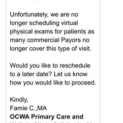
Images have been censored by Google after external flags. Re-wrote verbatim below so facts stay clear.
DIAGNOSIS SUBMITTED FOR INSURANCE APPROVAL:
[D25.0] Submucous Leiomyona of uterus
RAMANATHAN PER EPIC/MEDICAL RECORD:
Finding(s): 1. Small mobile uterus on exam under anesthesia. 2. Normal appearing liver, gallbladder, stomach on laparoscopy. Uterus with small fibroid. Normal right Fallopian tube. Normal bilateral ovaries. 3. On cystoscopy, normal appearing bladder with no injury or foreign body. Brisk bilateral ureteral jets. She had adenomyosis on pathology. No other endometriosis seen at time of surgery.
FINAL PATHOLOGY REPORT:
A) Uterus, cervix, left fallopian tube, total hysterectomy and left salpingectomy:
- Adenomyosis.
- Inactive endometrium, cervix and left fallopian tube, negative for neoplasm.
Specimenserus, with or without tubes and ovaries, other than neoplastic/prolapse, Uterus, cervi, let flopian tubes
Pre-op diagnosis:
Submucous leiomyoma of uterus [D25.0]
A. Uterus, with or without tubes and ovaries, other than neoplastic/prolapse.
Uterus, cervix, left fallopian tubes" is a 69 g, 7.5 x 4.6 x 3 cm uterus and cervix with attached left fallopian tube. Uterine serosa is tan-pink smooth and glistening. The anterior cervical margins of resection are inked blue, posterior black. The ectocervix is tan-pink and focally hemorrhagic around the patent 0.5 cm in diameter cervical os.
The endocervical canal and the cervical cut surfaces are normal. Endometrium is tan-pink flattened glistening measuring 0.1 cm in thickness. Uterus is serially sectioned the myometrium is tan-pink and rubbery and measures up to 2 cm in thickness. In the anterior wall there is a subendometrial 2 cm trabeculated focally cystic nodule consistent with adenomyosis. The remaining myometrium is unremarkable. No leiomyomata are identified.
The left fallopian tube measures 6.5 cm length is diameter ranges from 0.5 cm to 1 cm. Serosa is congested, smooth and glistening. There is a patent stellate lumen on cut surfaces
A1: Anterior cervix
A2: Posterior cervix
A3: Anterior comyometrium with subendometrial cystic lesion consistent with adenomyosis
A4: Posterior endomyometrium
A5: Cross-sections of left fallopian tube and is entire fimbriated end